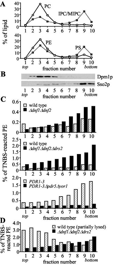
Figure 8.
Removal of Drs2p from Δdnf1Δdnf2 cells enhances cell surface exposure of endogenous PE. Wild-type and mutant cells were labeled with 10 mM TNBS as in Figure 7, lysed, and fractionated on sucrose step gradients. (A) Fractionation profiles of the different phospholipid species. (B) Fractionation profiles of plasma membrane (Sso2p) and ER (Dpm1p). (C and D) Distribution of TNBS-reacted PE, expressed as percentage of the total PE per gradient fraction. Lipid and protein profiles shown in A and B are from fractionated wild-type cells, but are representative for the profiles obtained with fractionated mutant cells. Results shown are from one experiment that was representative of two. The percentage of viable cells was 99.2 (wild-type), 89.9 (wild-type, partially lysed before TNBS labeling), 98.7 (Δdnf1Δdnf2), 93.8 (Δdnf1Δdnf2Δdrs2), 99.2 (PDR1–3), and 99.3 (PDR1–3Δpdr5Δyor1).

