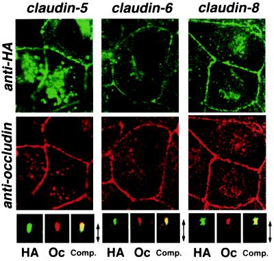
Figure 4.
Concentration of HA-tagged claudins at the occludin-positive tight junctions in MDCK transfectants. Confluent cultures of MDCK transfectants expressing HA-tagged claudin-5, -6, or -8 (claudin-5, claudin-6, claudin-8) were doubly stained with anti-HA mAb (anti-HA) and anti-occludin mAb (anti-occludin). Images were obtained at the focal plane of the most apical region of lateral membranes by confocal microscopy. HA-tagged claudins were coconcentrated with occludin at the level of TJs. As shown in the bottom panels, in computer-generated cross-sectional views, HA-claudins (HA) and occludin (Oc) were highly concentrated at the most apical portion of lateral membranes of MDCK cells, and overlaid images showed that HA-claudins were precisely colocalized with occludin (Comp.). The thickness of each cellular sheet is indicated by arrows. The same results were obtained from MDCK transfectants expressing HA-tagged claudin-3, -4, and -7 (data not shown).

