
Figure 3.
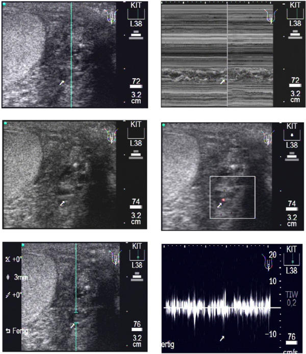
Longitudinal scan of a patient's left testis. Upper left: A medium sized para-testicular worm nest (arrow) is presented in this b-mode image. Upper right: The following m-mode section is positioned at the location of the largest diameter of the worm nest. The corresponding video image can be seen as Additional File 7: Movie3A.mpg. Middle left: The same worm nest as above. Middle right: The Colour Doppler-mode presents very few red signals as sign of less lymphatic fluid moved by the adult worms in this dilated lymphatic vessel. The corresponding video image can be seen as Additional File 8: Movie3B.mpg. Lower left: The same worm nest as above. The caliper for the PWD is positioned where the worm nest is located. Lower right: The Pulse Wave Doppler-mode confirms the medium sized worm nest by the irregular undulating band caused by the typical movements of the adult worms. The corresponding video image can be seen as Additional File 9: Movie3C.mpg.