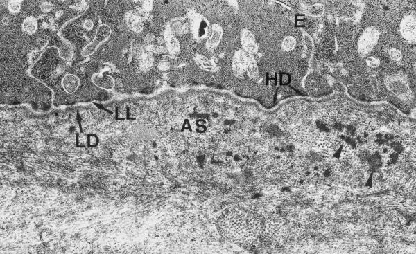
Figure 9.
Experimental cornea 3 months after the procedure. The basal part of epithelial cells demonstrates well-developed adhesive contacts. Hemidesmosomes (HD) have normal structure and regular distribution along basal lamina. The thickness and electron density of lamina densa (LD) and lamina lucida (LL) have returned back to normal values. The layer of anterior stroma (AS) contains aggregations of electrondense substance (arrowheads). The fibers of this layer have attained more regular distribution in comparison to 1-month post-op specimens. Electron microphotograph. Original magnification × 10 000.

