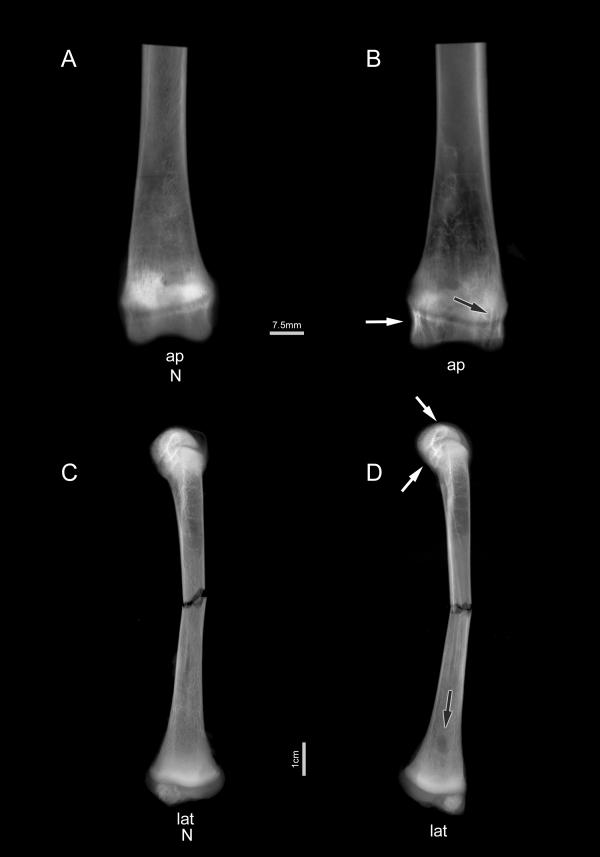
Figure 3.
Representative radiographs of the tibia at 8 weeks. Radiographs (A) and (B) show representative distal tibiae in anteroposterior (ap) position with growth plates still visible (horizontal bar = 7.5 mm). Bone from hCySH-treated animals (B) shows characteristic linear densities (white arrow) and spherical lucencies (black arrow) in cancellous bone in comparison to controls (A). Below are representative lateral (lat) radiologic views (vertical bar = 1 cm) of whole tibiae after loading to fracture. Here, a distinctive lucency (black arrow) is readily apparent in bone treated with hCySH (D) in comparison to control (C), but the trabecular elements of the secondary spongiosa seen in the anteroposterior radiographs are not as dramatic in these views. The metaphyseal lucency corresponds to an unmineralized avascular collagen 'plug' typical of chondrodysplasia. Note also the difference in rupture pattern.

