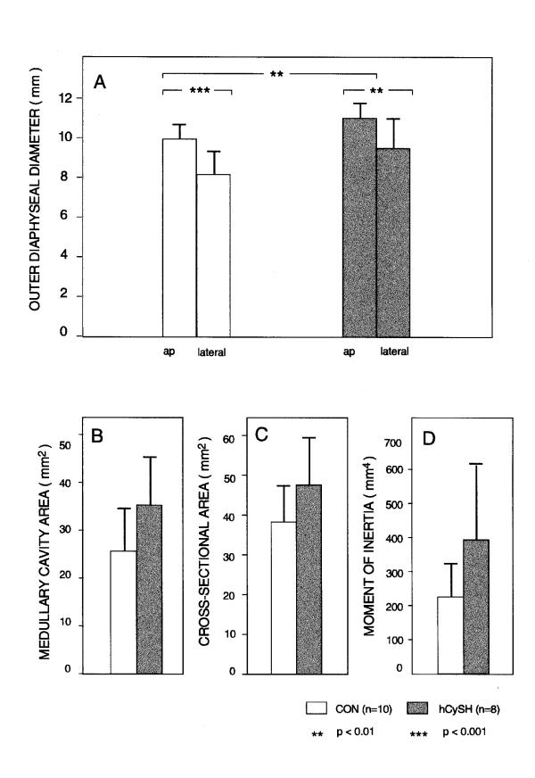
Figure 4.
Geometric characteristics and structural properties of the tibia at mid-shaft. In panel (A) are shown the anteroposterior (ap) and lateral outer diaphyseal diameters (mean " SD). AP diameters are significantly greater than lateral diameters in both groups (P < 0.01). The difference between groups is also significant for AP diameters. Panels (B), (C), and (D) show medullary cavity area (mm2), cross-sectional area (mm2), and moment of inertia (mm4), respectively. Dimensions for bone from hCySH-treated chicks was consistently greater.

