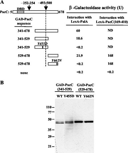
FIG. 3.
Two-hybrid interaction of PalA with PacC. Yeast strain CTY10-5d was used, and proteins were expressed from plasmids listed in Table 2. (A) GAD-PacC fusions contain the indicated PacC residues. The shaded bar indicates the DNA binding domain (DBD). Arrows mark the approximate position of the signaling-protease (∼493 to 500) (8) and processing-protease (∼252 to 254) (25) cleavage sites. Values are the average β-galactosidase activity of four transformants. Standard errors were <14%. In control experiments, GAD protein fusions did not interact with LexA (<0.4 U). ND, not determined. (B) Western analysis of protein extracts from transformants expressing LexA-PalA and the indicated GAD-PacC protein fusions which were detected with anti-HA antibodies. WT, wild type.

