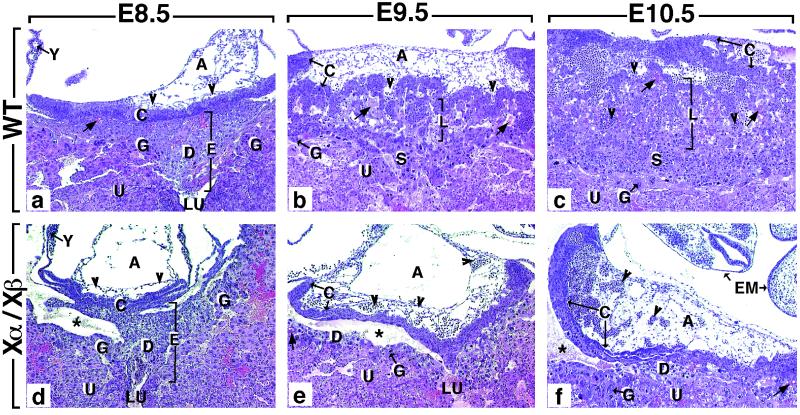
Figure 2.
Comparison of histological sections through the implantation site of wild-type (WT) (a–c) and RXRα−/−/RXRβ−/− (Xα/Xβ) mutants (d–f). (a) In E8.5 WT embryos, the polar extraembryonic ectoderm has generated the ectoplacental cone (E) and the chorionic (ectoplacental) plate (C). The ectoplacental cone penetrates into the uterine epithelium (U). It contains embryo-derived diploid and polyploid (giant) trophoblast cells (D and G, respectively), as well as maternal blood sinuses (arrows). The chorionic plate forms a solid wall of epithelial cells continuous with the ectoplacental cone and is fused to the mesoderm of the allantois (A), which contains extraembryonic blood vessels (arrowheads). (b) At E9.5, the placenta consists of four regions: (i) the chorionic plate (C), which is now traversed by allantoic capillaries (arrowheads); (ii) the labyrinthine zone (L), which is composed of strands of diploid trophoblast cells and of a network of extraembryonic capillaries interspersed with maternal blood sinuses; (iii) the spongiotrophoblast zone (S), in which only maternal blood circulates; and (iv) the giant cell zone (G). (c) At E10.5, the labyrinthine zone has enlarged markedly. (d–f) The size of the structures contributing to the mutant placenta increases between E8.5 and E10.5, but their differentiation is arrested at E8.5 (see the main text for further details). A, allantois; C, chorionic plate; D, diploid trophoblast; E, ectoplacental cone; EM, embryo; G, giant cell zone; L, labyrinthine zone; S, spongiotrophoblast zone; U, uterine epithelium; LU, uterine lumen; Y, yolk sac; arrowheads and arrow point to allantoic capillaries (containing nucleated erythrocytes) and to maternal blood sinuses, respectively. The asterisk indicates an artifactual tissue detachment generated during embedding. (×50.)

