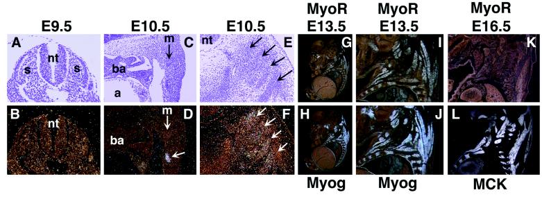
Figure 2.
Detection of MyoR and myogenin expression in developing skeletal muscle by in situ hybridization. Transcripts for MyoR were detected in transverse sections of E9.5 (B) and E10.5 (F) and in sagittal sections of E10.5 (D), E13.5 (G and I), and E16.5 (K) embryos. Expression of myogenin at E13.5 is illustrated in sagittal sections (H and J). Transcripts for MCK are shown in sagittal section of an E16.5 embryo (L). I and J show enlarged views of G and H, respectively. At E16.5, MyoR expression is down-regulated in contrast to the abundant expression of MCK. a, Atrium; ba, branchial arch; m, myotome; nt, neural tube; s, somite. The arrow in D shows a cluster of MyoR-expressing cells in body wall ventrolateral to the myotome (see text). Arrows in E and F mark differentiating axial skeletal muscle. (A and B, ×60; C–F, ×30; G and H, ×4; I and J, ×8; and K and L, ×5.)

