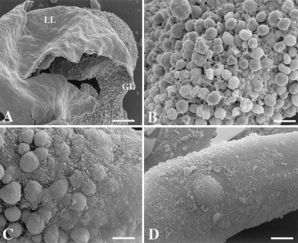
FIG. 2.
SEM of nontreated (A and B) or NTZ-treated (C and D) E. multilocularis metacestodes. (A and B) Control metacestodes cultured in vitro in the presence of DMSO (1:1,000) but in the absence of any drugs. Note that most cells exhibit an intact morphology. LL, laminated layer; GL, germinal layer. (C and D) Metacestodes cultured in vitro in the presence of 10 μg of NTZ/ml for 4 days (C) and 7 days (D). Substantial portions of the germinal layer already show massive signs of cellular destruction after 4 days of drug treatment but more clearly show massive signs of cellular destruction after 7 days of drug treatment and are detached from the laminated layer. Bars, 800 μm (A), 280 μm (B), 240 μm (D), and 320 μm (E). Similar results were obtained for parasites treated with 5 μg of NTZ/ml or TIZ and TIZ gluc (data not shown).

