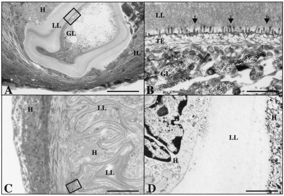
FIG. 5.
Light microscopy (A and C) and corresponding TEM (B and D) of tissue recovered from mice which had been infected with DMSO-treated metacestodes (A and B) or NTZ-treated metacestodes (C and D). Note the presence of viable parasite in panel A, and a region similar to the one framed in panel A is shown by TEM in panel B. (C) The laminated layer is completely encapsulated by host tissue, and a region similar to the one framed in panel C is shown by TEM in panel D. Note that no germinal layer is visible in tissue recovered from mice infected with NTZ-treated metacestodes. Bars, 120 μm (A), 4.2 μm (B), 60 μm (C), and 1.2 μm (D). GL, germinal layer; TE, tegument; LL, laminated layer; H, host tissue.

