Figure 3.
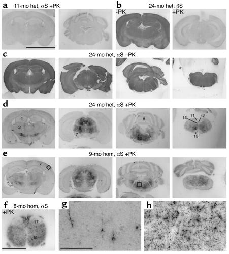
Age-dependent formation of PK-resistant αS in transgenic mice. (a) αS PK-PET blots of an 11-month-old heterozygous (het) mouse showed no PK-resistant αS. (b) βS PK-PET blots of a 24-month-old heterozygous mouse showed no PK-resistant βS (+PK) despite strong staining of PK-labile βS (–PK). (c and d) PK-PET blots of a 24-month-old heterozygous mouse before (c) and after (d) PK treatment showed specific staining of PK-resistant αS only in specific brain regions, although transgenic αS was expressed throughout the brain. (e) The same pathology was already seen on αS PK-PET blots of a 9-month-old homozygous (hom) mouse. (f) αS PK-PET blot of the SC of an 8-month-old homozygous mouse. (g and h) Higher magnifications of the areas boxed in e, namely cortex (g) and pontine reticular field (h). Scale bar in a, 5 mm (a–e); in f, 1 mm; in g, 200 μm (g and h). Numbers in d and f: 1, hippocampus; 2, thalamus; 3, zona incerta; 4, aqueduct and periaqueductal gray; 5, superior colliculus; 6, mesencephalic nucleus; 7, SN; 8, cerebellum; 9, facial nerve root; 10, pontine zona reticulata; 11, nucleus of solitary tract; 12, aqueduct; 13, hypoglossal nucleus; 14, medullary zona reticulata; 15, pyramidal tract; 16, ventral horn; 17, dorsal horn; 18, dorsal columns.

