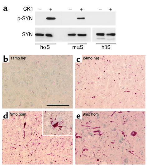
Figure 5.
Pathological αS hyperphosphorylation in transgenic mice. (a) Human αS, mouse αS, and human βS were incubated without (–) or with (+) casein kinase 1 (CK1), and 20 ng synuclein aliquots were Western-probed with phosphospecific anti-αS (p-SYN, upper panel), MC42 anti-αS, and 6485 anti-βS, respectively (SYN, lower panels). (b–e) Immunohistochemistry with phosphospecific anti-αS of the pontine reticular field (b–d) and ventral horn of the SC (e) from heterozygous mice aged 11 months (b) and 24 months (c) as well as from a homozygous mouse aged 9 months (d and e). Inset in d highlights phospho-αS–containing neuronal cytosolic inclusions and neuritic swellings. Scale bar, 100 μm in b–e, 50 μm in inset.

