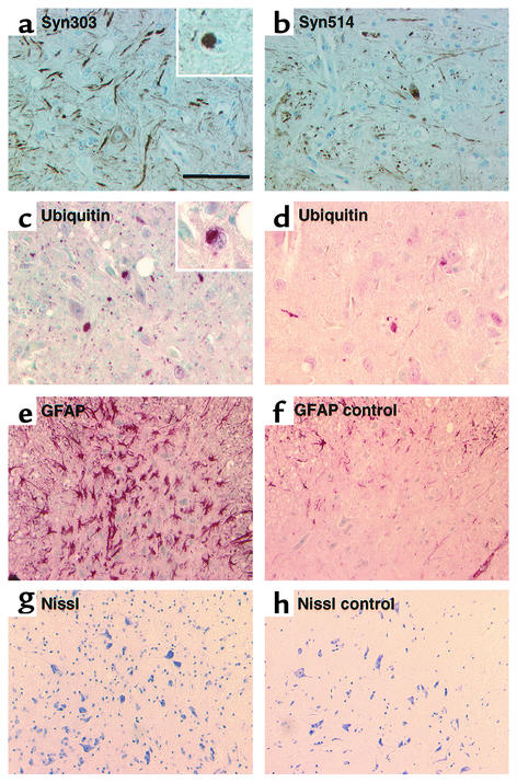
Figure 6.
Pathology in severely affected transgenic mouse brain regions. (a and b) Antibodies Syn303 (a) and Syn514 (b) against oxidatively modified αS stained numerous pathological neuritic profiles and occasional LB-like neuronal inclusions, one of which is magnified in the inset in a. (c and d) Anti-ubiquitin immunostaining showed some neuritic and cell body inclusions (inset in c). a–c are from the pontine reticular nuclei, and d is from the ventral horn of the SC. (e and f) Immunostaining of glial fibrillary acidic protein (GFAP) revealed prominent astrocytic gliosis in the most affected regions (here the ventral horn of the SC) in a motor-impaired homozygous (Thy1)-αS mouse (e) compared with a nontransgenic control mouse (f). (g and h) Nissl staining of the identical area of ventral horn revealed numerous motor spinal neurons in transgenic (g) and control (h) mice. Scale bar, 100 μm in a–d, 50 μm in inserts in a and c, 200 μm in e–h.

