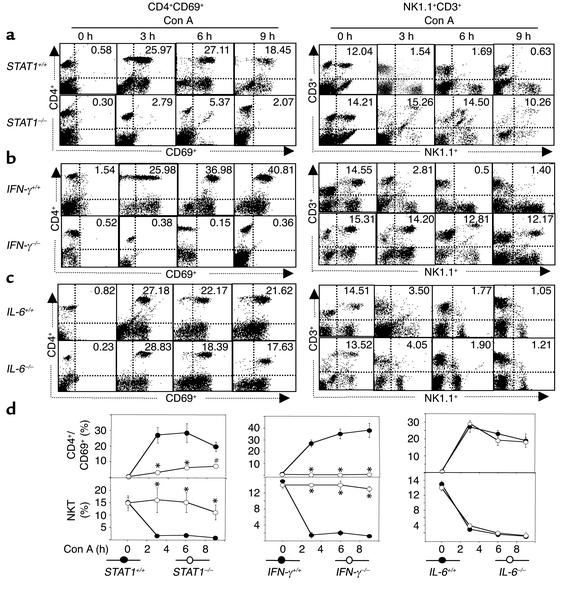
Figure 6.
Con A injection–mediated activation of CD4+ and NK T cells is abolished in STAT1–/– and IFN-γ–/– but not in IL-6–/– mice. Wild-type and knockout mice were injected with Con A for 3, 6, and 9 hours. Hepatic lymphocytes were isolated. The surface of CD4+CD69+ or NK1.1+CD3+ was analyzed by flow cytometry. The flow cytometric analysis is representative of three independent experiments. The upper-right quadrant in each panel shows CD3+CD69+ or NK1.1+CD3+ double-positive cells (percentage of the total hepatic lymphocytes). Values are shown in d as means ± SEM from three mice at each time point. *P < 0.001 and #P < 0.01 vs. corresponding Con A–treated wild-type groups at the same time points.

