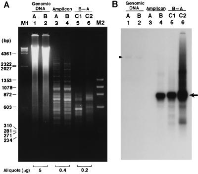
Figure 2.
Actual course of AP-RDA using the AP-3 primer. (A) Ethidium bromide staining. Electrophoresis was performed at 100 V for 3 hours. (B) Probed with a polymorphic clone, AP3-G7. Lanes: 1 and 2, BamHI-digested genomic DNAs from ACI and BUF rats; 3 and 4, amplicons of ACI and BUF; 5 and 6, C1 and C2 products of AP-RDA using AP-amplicon from BUF as the tester and that from ACI as the driver. M1 and M2, λ/HindIII and φX174/HaeIII DNA size markers. The arrow on the right shows a polymorphic band detected by AP3-G7 clone. The arrowhead shows hybridization of the genomic DNAs of ACI and BUF with this probe.

