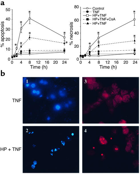
Figure 2.
Mitochondrial GSH depletion sensitizes hepatocytes to TNF-α. (a) Cultured hepatocytes were exposed to TNF-α (280 ng/ml, solid line; 25 ng/ml, dashed line) for various periods of time with or without HP preincubation to deplete the mGSH levels. Cell death was determined by double staining with Hoechst 33258 and propidium iodide to detect apoptotic and necrotic cells, respectively. At least 250 cells in six different high-power fields were counted and expressed as a percentage of total cells (please note the scale difference). HP alone did not affect cell survival. Results are expressed as means ± SD (n = 4 experiments). *P < 0.05 versus control. (b) Representative fluorescent micrographs of control hepatocytes exposed to TNF-α (panels 1 and 3) or HP-pretreated hepatocytes (panels 2 and 4). Blue fragmented nuclei (panel 2) represent apoptotic cells, whereas red nuclei (panel 4) are indicative of necrotic cells.

