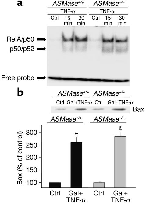
Figure 9.
NF-κB activation and mitochondrial Bax translocation in mouse hepatocytes. (a) Nuclear extracts from ASMase+/+ and ASMase–/– hepatocytes after TNF-α exposure were prepared and used for NF-κB activation using a consensus oligonucleotide. (b) Hepatocytes were treated with galactosamine plus TNF-α (280 ng/ml) for 16 hours. Cells were then fractionated to prepare mitochondria as described in the Methods. Bax levels in the mitochondrial fraction were analyzed by Western blotting and quantitated by densitometry. Results are expressed as means ± SD (n = 4 independent experiments). *P < 0.05 versus control. Gal + TNF-α, galactosamine plus TNF-α.

