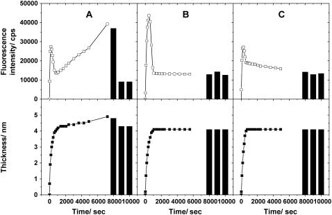
FIGURE 4.
Kinetic observation of vesicle fusion by SPR-SPFS. The lipid layer thickness observed by SPR (lower frame) and the fluorescence intensity observed by SPFS (upper frame) were plotted versus incubation time for vesicles of (a) POPC, (b) POPC/POPS (4:1), and (c) POPC/DOTAP (4:1). A trace amount of DiD (10−4 mol/mol) was added to all samples. The total concentration of vesicle suspensions was 0.1 mM. The solid columns indicate values after successive rinsings with the buffer solution, Milli-Q water, and the buffer solution, respectively (from left to right). Milli-Q water and buffer solution have different refractive indices. This difference was incorporated in the SPR curve fitting to evaluate the adsorbed layer thickness.

