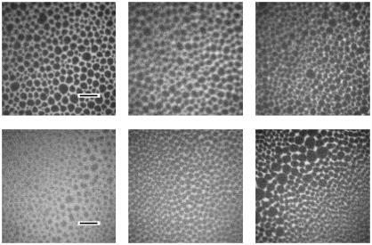
FIGURE 2.
Fluorescence light micrographs of surfactant model systems spread on a film balance: (top) DPPC/DPPG/SP-B (80:20:0.4 mol %); (bottom) DPPC/d62DPPG/SP-B (80:20:0.4 mol %). The pictures are taken at 5 mN/m (left), 30 mN/m (middle), and 50 mN/m (right). The length of the black scalebar is 20 μm.

