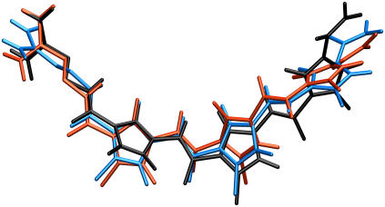
FIGURE 5.
Relative motion of distamycin inside the DNA minor groove. Three representative snapshots of distamycin from the 10-ns simulation of the distamycin-DNA complex are shown superimposed after least-square fitting: initial conformation (black), and conformations corresponding to the first (blue) and second (red) rapid increase in configurational entropy (see arrows in Figs. 3 and 6).

