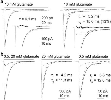
FIGURE 2.
Glutamate-induced desensitization shows multiple kinetic components that do not depend on receptor occupancy. (a) Current evoked by 100-ms applications of 10 mM glutamate. Left and right panels show one- and two-exponential fits (solid curves) to the decays. The residuals are also shown (res; obtained by subtracting the data from the fit). Insets are the same currents and fits on an expanded timescale (dashed lines show individual components). (b) Currents evoked by sustained applications of two different concentrations of glutamate (left). In the panels to the right, the individual currents were scaled to have the same peak amplitude. The individual components obtained from the biexponential fits to the decays are shown as dashed curves.

