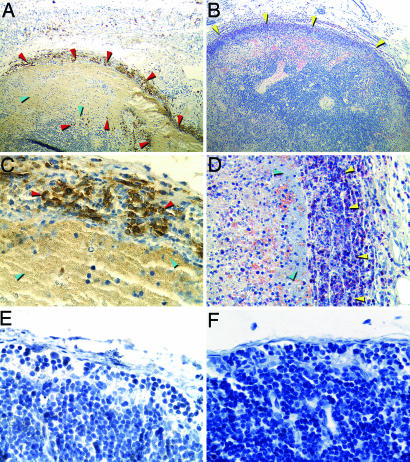
Fig. 3.
iNOS expression by PMNs in the bubo. Shown are sections of a bubo (A–D) or uninfected lymph node (E and F) stained to detect iNOS (A, C, and E) or PMNs (B, D, and F; yellow arrowheads). PMNs producing iNOS (dark brown) are indicated by red arrowheads. Masses of extracellular bacteria adjacent to PMNs are indicated by blue arrowheads. (Magnification: A and B, ×100; D, ×400; C, E, and F, ×600.)

