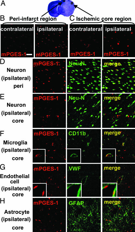
Fig. 2.
Induction of mPGES-1 in neurons, microglia, and endothelial cells in the cortex after ischemia. (A) The predesignated areas in the peri-infarct and ischemic core regions of the postischemic cortex for microscopic examination. (B and C) The immunostaining of mPGES-1 in the peri-infarct region (B) and ischemic core region (C) of ipsilateral postischemic cortex and contralateral cortex. (D–H) The double-immunostaining of mPGES-1 (red) and cell-type-specific marker proteins (green) in the peri-infarct (D) and core (E–H) regions of the cortex. Neurons (D and E), microglia (F), endothelial cells (G), and astrocytes (H) were recognized by antibodies for Neu-N, CD11b, von Willebrand factor (VWF), and GFAP, respectively. Insets show high magnification of each staining. The photographs shown are representative examples from three separate experiments. (Scale bars: main images, 40 μm; Insets, 10 μm.)

