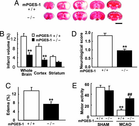
Fig. 4.
Deletion of the mPGES-1 resulted in marked amelioration of the infarction, edema, and behavioral neurological dysfunctions observed after ischemia. (A) Representative 2,3,5-triphenyltetrazolium chloride (TTC)-stained coronal sections at −2, 0, +2, +4, and +8 mm from the bregma of the mPGES-1 KO (−/−) and WT (+/+) mouse. (Scale bar: 5 mm.) (B) The volume of infarcted brain tissue 24 h after ischemia was estimated and expressed as a percentage of the corrected tissue volume. n = 10 animals per group; ∗∗, P < 0.01 vs. WT mice. (C) The corrected edema percentage in the mPGES-1 KO (−/−) and WT (+/+) mice. n = 10 animals per group; ∗∗, P < 0.01 vs. WT mice. (D) Improvement of neurological dysfunction in mPGES-1 KO mice. The neurological score was measured 24 h after ischemia. n = 21–22 animals per group; ∗∗, P < 0.01 vs. WT mice. (E) The motor activity of MCAO or sham-operated (SHAM) mice. n = 9–10 animals for SHAM and n = 19–20 animals for MCAO; ∗∗, P < 0.01 vs. SHAM WT mice; ##, P < 0.01 vs. MCAO-treated WT mice.

