Fig. 5.
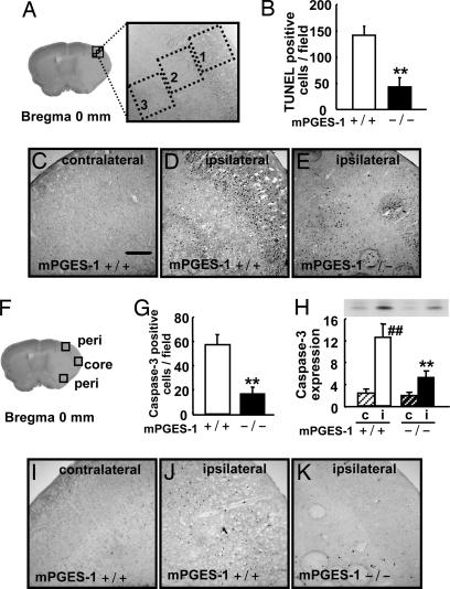
Deletion of the mPGES-1 gene resulted in a marked decrease in the apoptotic neuronal death in the postischemic cortex. (A) Three predesignated areas in the penumbra of the postischemic cortex for counting TUNEL-positive cells. (B) The average number of TUNEL-positive cells per unit area (A) of mPGES-1 KO (−/−) and WT (+/+) mice. n = 7 animals per group; ∗∗, P < 0.01 vs. WT mice. (C–E) Representative results of TUNEL staining in the contralateral or ipsilateral cortex of the mPGES-1 KO and WT mouse. (Scale bar: 100 μm.) (F) Three predesignated areas in peri-infarct (peri) and core regions for counting caspase-3-positive cells in the cortex. (G) The average number of caspase-3-positive cells per unit area (F) after transient ischemia. n = 7 animals per group; ∗∗, P < 0.01 vs. WT mice. (H) The tissue lysates from the contralateral (c) or ipsilateral (i) cortex were subjected to Western blot analysis for caspase-3. The densities of immunoblots were measured. n = 5 animals per group; ##, P < 0.01 vs. contralateral cortex. (I–K) Representative caspase-3 immunostaining in the cortex.

