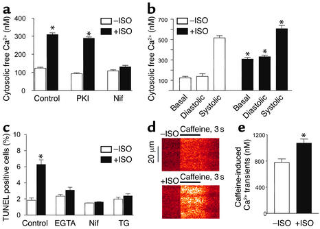
Figure 4.
PKA-independent increase in intracellular Ca2+ is essential for the β1AR apoptotic effect. After β1β2 DKO myocytes were infected by Adv-β1AR, cells were incubated with designated reagents for 1 hour, then ISO (1 μM) was added and cells were incubated for another 3–6 hours (a, b, d, and e) or 24 hours (c). (a) Prolonged β1AR stimulation elevated basal intracellular free Ca2+ in unpaced cardiac myocytes. This effect was blocked by the L-type Ca2+ channel antagonist nifedipine (1 μM), but not the PKA inhibitor PKI (5 μM). *P < 0.01 vs. ISO-untreated groups and those pretreated by nifedipine (n = 20–35 cells from six hearts). (b) Intracellular Ca2+ transients were measured in a subset of cells electrically paced at 0.5 Hz for at least 10 minutes in the absence (n = 29 cells from four hearts) and presence (n = 22 cells from four hearts) of sustained β1AR stimulation by ISO. *P < 0.05 vs. ISO-untreated myocytes. (c) Effects of nifedipine, EGTA-AM (1 μM), or the SR ATPase inhibitor thapsigargin (1 μM) on β1AR-induced increase in TUNEL-positive cells. *P < 0.01 vs. ISO-untreated myocytes and those pretreated with EGTA-AM, nifedipine, or thapsigargin (n = 4–8). (d) Representative confocal linescan images of caffeine-elicited SR Ca2+ release in ISO-treated (1 μM, 3 hours, bottom) and untreated cells (top). The x axis shows the time courses for caffeine treatment, and the y axis shows the spatial profiles of Ca2+ transients along a scan line inside the cell. (e) Average amplitude of caffeine-elicited Ca2+ transients in ISO-treated or untreated group. *P < 0.01 vs. ISO-untreated myocytes. n = 25–30 cells from six hearts in each group. Nif, nifedipine; TG, thapsigargin.

