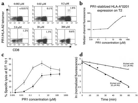
Figure 1.
Lower doses of PR1 peptide induce CTLs with higher intensity PR1/HLA-A2 tetramer staining that correlates with TCR avidity and inversely with effector function threshold. (a) PBMCs collected from healthy HLA-A2.1+ donors were stimulated weekly with PR1 peptide-pulsed T2 cells at the peptide concentrations indicated above each FACS plot. After 4 weeks, resulting cultures were stained with CD8 (FITC) Ab and PR1/HLA-A2 tetramer, and the percentage of CD8+ cells that stain with tetramer are noted within each FACS plot. (b) Surface HLA-A2 expression on T2 cells increases linearly with increasing concentration of PR1 peptide from 2 μM and 200 μM. T2 cells were incubated with PR1 peptide at the concentrations shown and surface HLA-A2 expression was measured by flow cytometry. (c) CTLs elicited with PR1 at 0.2 μM (open circles) or 20 μM (filled squares) PR1 were incubated for 4 hours at 37°C with PR1 peptide-pulsed T2 cells at the indicated peptide concentrations at an effector/target (E/T) ratio of 10:1 (adjusted based on the number of tetramer-positive CTLs), and percentage of specific lysis was determined. (d) Tetramer decay (t1/2) was determined to be 58 minutes and 19 minutes by plotting normalized antigen-specific fluorescence at the indicated time points for 28-day-old PR1/HLA-A2 tetramer-stained CTLs elicited with 0.2 μM (open circles) or 20 μM PR1 (filled squares), respectively. Dissociation kinetics of PR1/HLA-A2 tetramer staining were determined at 4°C in the presence of saturating concentrations of BB7.2 Ab to prevent rebinding of tetramer and in the presence of PI (1 μg/ml) to eliminate dead cells from the FACS gate.

