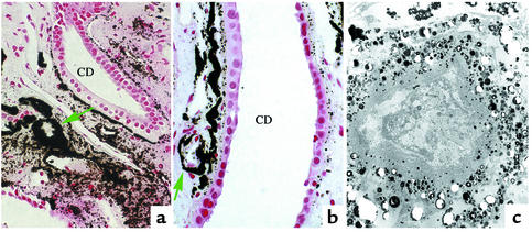
Figure 4.
Accumulation of interstitial crystal deposits. In (a) and (b), extensive accumulation of crystalline deposition (green arrows) is shown around a few loops of Henle and nearby vascular bundles, resulting in the formation of incomplete to complete cuffs (indicated by circles) of crystalline material in the papillary tissue of a CaOx patient. Note the accumulation of crystal material in the basement membrane of nearby collecting duct and the normal appearance of the collecting duct cells. In (c), an electron micrograph shows a dense accumulation of crystalline material around a loop of Henle that appears to be necrotic. Magnification, ×900 (a); ×1,600 (b); ×400 (c). CD, collecting duct.

