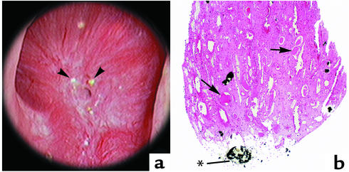
Figure 6.
Endoscopic and histologic images of Randall’s plaques in intestinal bypass patients. In (a), an example of a papilla from an intestinal-bypass stone former that was video recorded at the time of the mapping is shown. Distinct sites of Randall’s plaque material are not found on the papilla of the intestinal-bypass patient; instead, several nodular-appearing structures (arrowheads) were noted near the opening of the ducts of Bellini. In (b), a low magnification light microscopic image of a papillary biopsy specimen from an intestinal-bypass patient is shown. Crystal deposition was only found in the lumens of a few collecting ducts as far down as the ducts of Bellini (*). A large site of crystal material was seen in a duct of Bellini. No other sites of deposits were noted. Note dilated collecting ducts (arrows) with cast material and regions of fibrosis around crystal-deposit–filled collecting ducts. Magnification, ×100 (b).

