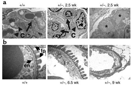
Figure 3.
Heterozygous VEGF-loxP+/–,Neph-Cre+/– mice demonstrate endotheliosis and loss of fenestrations. (a) At 2.5 weeks of age, wild-type glomerular capillary loops (c) are open and contain numerous red blood cells. In contrast, podocyte-specific VEGF-A heterozygotes (+/–) demonstrate bloodless glomeruli, and the capillary loops are filled with swollen endothelial cells, demonstrating endotheliosis, the classic renal lesion of preeclampsia. In addition, large subendothelial hyaline deposits (*) can be seen. (b) At 6.5 weeks of age, wild-type filtration barriers (+/+) are characterized by fenestrated endothelial cells (en) and well-formed podocyte foot processes (fp). In the podocyte-specific heterozygotes (+/–), the fenestrations are lost at 6.5 weeks of age, and by 9 weeks of age, the endothelial cells appear necrotic and no podocyte foot processes can be identified.

