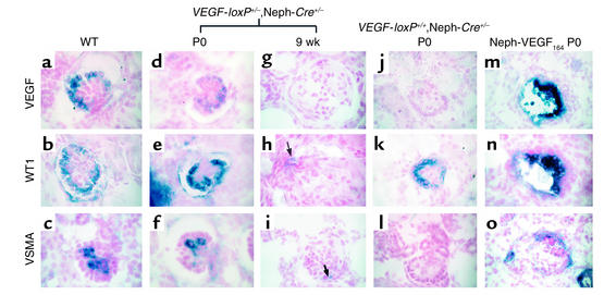
Figure 4.
Digoxigenin-labeled in situ analysis of wild-type and mutant glomeruli. (a–c) Capillary loop–stage glomeruli from a newborn VEGF-loxP–/–,Neph-Cre+/– control mouse demonstrate expression of VEGF-A and WT1 in podocytes, while VSMA is expressed in mesangial cells, which are found inside the glomerulus and are required to support the capillary structure. (d–f) At birth (P0), capillary loop–stage glomeruli from a heterozygous VEGF-loxP+/–,Neph-Cre+/– mouse demonstrate normal levels of expression of WT1 and VSMA, while VEGF expression is consistently reduced at the mRNA level compared with the wild-type controls. (g–i) By 9 weeks of age, the heterozygous VEGF mice are clinically unwell. At this time, most glomeruli demonstrate a complete absence of markers of podocyte differentiation (i.e., no VEGF or WT1; both are absent). In h, a single WT1-positive cell can be identified (arrow). (i) VSMA is not usually present in glomeruli at 9 weeks; however, occasional VSMA-positive cells can also be identified and likely represent “activated” mesangial cells (arrow). (j–l) In the null VEGF-loxP+/+,Neph-Cre+/– glomeruli at birth, no VEGF is seen in glomeruli as predicted due to podocyte-specific excision of both VEGF alleles. WT1 is present in differentiated podocytes. In contrast, VSMA is absent, demonstrating a defect in migration and/or differentiation of mesangial cells into the glomerulus. (m–o) In the nephrin–VEGF-164 mouse, both VEGF and WT1 are expressed in podocytes present within collapsed glomeruli. VEGF is markedly upregulated. In addition, VSMA and mesangial cells are present but appear to surround the collapsed glomerulus in a crescent shape. Magnification: ×350.

