Figure 6.
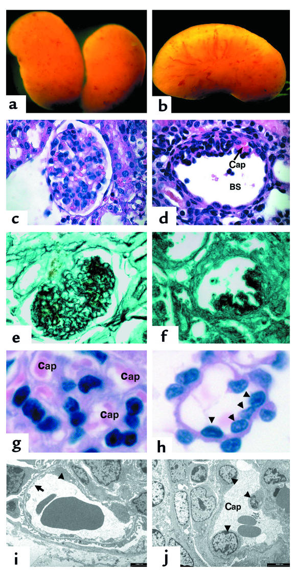
Mice that overexpress the 164 isoform of VEGF-A in their podocytes develop collapsing glomerulopathy. (a and b) Whole-mount images of VEGF-overexpressing kidneys at 5 days. The kidneys demonstrate many surface hemorrhages. (c) A glomerulus stained with H&E from a wild-type littermate. (d) A glomerulus from a transgenic VEGF-overexpressing mouse demonstrates global collapse of the capillary tuft toward the vascular pole of the glomerulus. A single patent capillary loop that appears dilated is identified (Cap). In addition, Bowman’s space (BS) is enlarged. (e) A 5-day-old wild-type glomerulus is stained with silver methenamine that recognizes basement membranes (black). Note the intricate pattern of GBM that lines the capillary loops between endothelial cells and podocytes. (f) In contrast, a transgenic glomerulus demonstrates complete collapse of the capillary network. (g) A high-power view of the capillary loops (Cap) in a wild-type glomerulus. (h) In contrast, the few patent capillary loops identified at 5 days of age in the transgenic mice demonstrate increased diameter and multiple endothelial cell nuclei (arrowheads). (i) A wild-type capillary loop at 5 days of age. Note the fenestrated endothelium (arrow). Although a portion of an endothelial cell body is identified (arrowhead), glomerular endothelial cell nuclei are difficult to find on EM sections. (j) In a transgenic patent capillary loop at 5 days of age, three endothelial cell nuclei are easily identified (arrowheads). Magnification in a and b: ×60; in c–f: ×225; in g and h: ×1,000. In i, bar = 2,000 nm; in j, bar = 5,000 nm.

