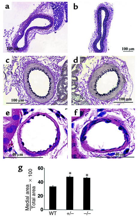
Figure 2.
Structures of aorta and renal vasculature in wild-type and RGS2-deficient mice. Histological analysis (hematoxylin-and-eosin or Verhoeff–Van Gieson elastin staining) of wild-type (a, c, and e) and rgs2–/– (b, d, and f) mice. Aorta (a and b), renal interlobular arteries (c and d), and nonelastic arterioles of the renal cortex (e and f) are shown. (g) Morphometric analysis of relative medial thickness (medial area/total area) of renal resistance vessels with diameters of 50–100 μm from wild-type, rgs2+/–, and rgs2–/– mice. Relative medial thickness of rgs2 mutants differed significantly (*P < 0.0001) from that of wild-type mice.

