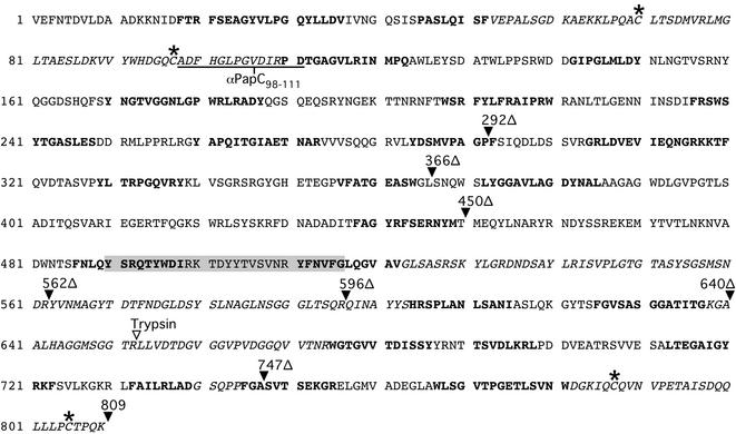
FIG. 1.
Sequence of mature PapC. Boldface lettering indicates TM β-strands predicted by computer modeling. Italic lettering indicates predicted loop regions experimentally localized to the periplasm. The conserved cysteine pairs are labeled with a star, the periplasmic epitope recognized by the αPapC98-111 antibody is underlined, and the periplasmic trypsin cleavage site is indicated by an inverted open triangle. The positions of the His-tagged C-terminal truncations are indicated, as well as the His tag appended to full-length PapC (both shown by inverted filled triangles). The residues boxed in gray (490 to 516) represent the corrected PapC sequence.

