Figure 2.
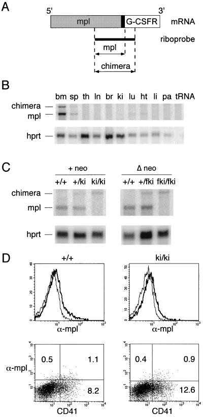
Expression of chimeric mpl/G-CSFR. (A) Ribonuclease protection assay. Position of riboprobe used to distinguish wild-type from chimeric mpl/G-CSFR mRNA. Solid box, transmembrane domain. Arrows, length of protected fragments for the wild-type mpl and chimeric transcripts. (B) Expression of wild-type and chimeric mRNA in tissues of heterozygous +/ki mice: bm, bone marrow; sp, spleen; th, thymus; ln, mesenteric lymph node; br, brain; ki, kidney; lu, lung; ht, heart; li, liver; pa, pancreas. A riboprobe for mouse hypoxanthine phosphoribosyltransferase (hprt) was used as an internal control for RNA loading. (C) Expression of chimeric mRNA in spleens of ki and wild-type mice. On the left is the original mouse strain containing the neo cassette (+ neo); on the right is the mouse strain after germ-line excision of the plox-neo gene (Δ neo); fki, the floxed ki allele. (D) Flow cytometric analysis of Percoll-fractionated bone marrow cells. In the top row is shown the biotinylated anti-mpl with streptavidin-PE (thick line) versus isotype control (thin line). In the bottom row is two-color staining with anti-mpl/streptavidin-PE and fluorescein isothiocyanate-labeled anti-CD41. Numbers indicate percentages of cells in each quadrant.

