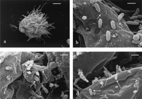
FIG. 1.
Scanning electron microscope views of B. pseudomallei and A. astronyxis coculture. (a) A. astronyxis trophozoite prior to inoculation with B. pseudomallei NCTC 13177. Scale bar = 10 μm. (b) Trophozoite surface covered with adherent B. pseudomallei NCTC 13177 cells, some of whose flagella (arrows) are visible. Scale bar = 2 μm. (c) B. pseudomallei NCTC 13177 cells (arrows) tucked in folds of the trophozoite surface during phagocytosis. Scale bar = 2 μm. (d) B. pseudomallei NCTC 13177 cells (arrows) remaining on the external trophozoite surface showing end-on adherence. Scale bar = 2 μm.

