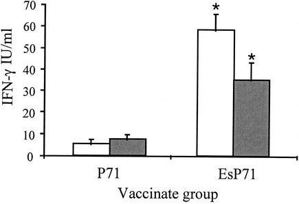
FIG. 2.
IFN-γ levels in splenocyte supernatants from P71 and EsP71 vaccinates following stimulation with P71. Mouse splenocytes from P71 and EsP71 vaccinates were seeded at a concentration of 5 × 10⁶ cells per well and were stimulated in vitro with 10 μg of P71 antigen/ml for 72 h. The IFN-γ levels were determined in the supernatant fractions by ELISA. Data represent means plus standard errors of two experiments with triplicate wells in each experiment. White bar, day 32; gray bar, day 42. An asterisk indicates that the value was significantly different (P < 0.05) from the values for other vaccinate groups for the same time point. Splenic lymphocytes from control VR1020-vaccinated mice did not respond to the P71 antigen (data not shown).

