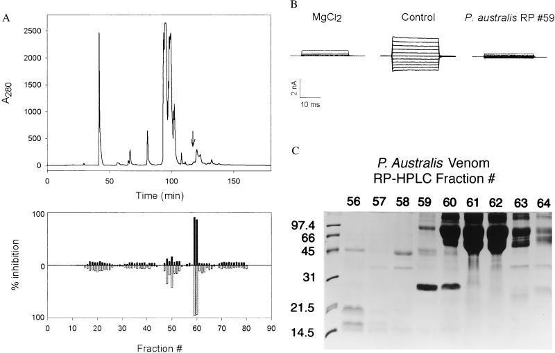
Figure 1.
(A) RP-HPLC separation of PsTx from P. australis venom. The components in 100 mg of venom were separated on a C5 column by using a water/acetonitrile gradient as described. (Upper) The column profile at 280 nm. Ten-milliliter fractions were dried and redissolved in 1 ml of water. After a 1:30 dilution into control buffer, the fractions were applied to excised outside-out patches expressing the olfactory CNG channel α-subunit. (Lower) Histogram depicts the fractional inhibition of current at +80 mV (upward, black fill) and at −80 mV (downward, gray fill). The PsTx eluted at 118 min. (indicated by an arrow, Upper); the corresponding inhibitory activity was found in fractions 59 and 60. These data suggest that PsTx is a minor component of P. australis venom, perhaps <0.1%. (B) Block of CNG channel currents by 10 mM Mg2+ and crude PsTx (reversed-phase fraction 59). CNG channel currents in excised outside-out patches were evoked by 20-ms steps from a holding potential of mV to a voltage ranging from −100 to +100 mV in 20-mV steps. Shown are current families in control solution and in the presence of 10 mM MgCl2 and PsTx applied to the extracellular face of the membrane. (C) SDS/PAGE analysis of reversed-phase fractions 56–64. A 10-μl sample of each reconstituted fraction was applied to a SDS-15% polyacrylamide gel. After separation, the gel was stained with Coomassie brilliant blue R250. Inhibitory activity was strongly correlated with the presence of a 24-kDa protein in fractions 59 and 60.

