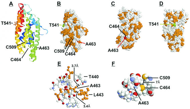
FIG. 11.
THREADER three-dimensional model of the IEP86 C-terminal fragment. Three-dimensional structures were predicted by using THREADER version 2.5 (17). (A) Shown is the ribbon structure colored from blue to red in the direction from the N terminus to the C terminus. Amino acids 463, 464, 509, and 541 are highlighted as ball-and-stick models. (B, C, and D) Shown are space-filling models where hydrophobic residues are colored in orange, T541 is shown in green, C464 and C509 are shown in black, and A463 is shown in red. (E) Shown are amino acids located 4 Å or less from A463. Hydrophobic residues are orange. (F) Shown are amino acids located 5 Å or less from C464.

