FIG. 6.
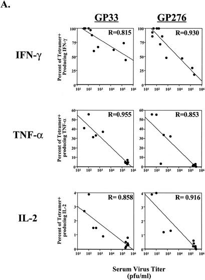
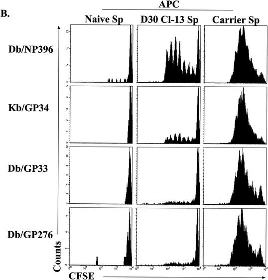
Correlation of antigen load to functional impairment of virus-specific CD8 T cells. (A) Virus in serum was quantitated by plaque assay, and the level of virus was plotted against cytokine responses. Top row, the percentage of tetramer-positive (tetramer+) cells producing IFN-γ in chronically infected mice (1 to 2 months p.i.) is plotted against viral load. Middle and bottom rows, the percentage of tetramer-positive cells that can synthesize TNF-α or IL-2 at day 15 after Cl-13 infection is plotted against the viral load. Lines indicate the linear regression best fit. (B) Splenocytes from uninfected mice (Naive Sp; left column), LCMV Cl-13-infected mice (day 30 p.i.; D30 Cl-13 Sp, middle column), or LCMV carrier mice (Carrier Sp, right column) were depleted of CD8 T cells and used as APC to stimulate CFSE-labeled, purified memory CD8 T cells from LCMV Armstrong-immune mice (>30 days p.i.). Proliferation was assessed after 60 h of coincubation by determining loss of CFSE fluorescence in the four tetramer-positive populations indicated on the left. Similar results were observed when APC were not depleted of CD8 T cells (data not shown).


