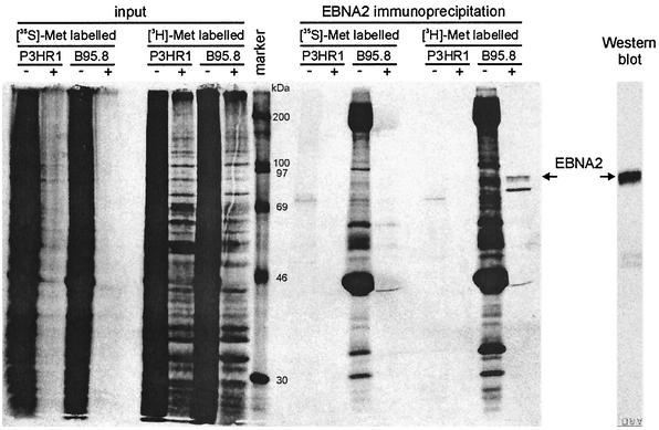
FIG. 2.
In vivo methylation of EBNA2. B95.8 cells (EBNA2 positive) and P3HR1-1 cells (EBNA2 negative) were labeled in vivo with either [35S]methionine or [3H-methyl]methionine in the presence (+) or absence (−) of the protein synthesis inhibitors cycloheximide and chloramphenicol. The cell extracts were subjected to immunoprecipitation with the EBNA2-specific MAb R3 (14) and analyzed by SDS-PAGE and autoradiography (lanes designated “EBNA2 immunoprecipitation”). The unprecipitated cell extracts (lanes designated “input”) were also analyzed to demonstrate that protein de novo synthesis was efficiently inhibited. The rightmost lane, designated “Western blot,” shows that MAb R3 efficiently precipitated the EBNA2 protein. Arrows indicate the positions of the precipitated EBNA2 protein; the lane designated “marker” shows the positions of coelectrophoresed 14C-labeled molecular mass marker proteins (Amersham).

