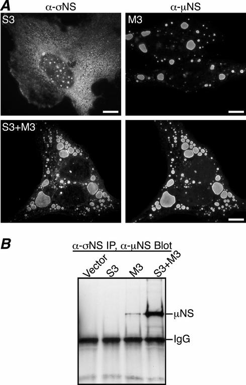
FIG. 2.
Colocalization and co-IP of σNS and μNS in transfected CV-1 cells. (A) IF microscopy of CV-1 cells at 18 h p.t. with pCI-S3 (top left), pCI-M3 (top right), or both pCI-S3 and pCI-M3 (bottom row). σNS was visualized by immunostaining with σNS-specific mouse MAb 3E10 (5) followed by Alexa 488-conjugated anti-mouse IgG (left column). μNS was detected by immunostaining with Texas Red-conjugated μNS-specific polyclonal antibodies (9) (right column). Scale bars, 10 μm. (B) CV-1 cells transfected with pCI-neo (Vector), pCI-S3, pCI-M3, or both pCI-S3 and pCI-M3 were lysed in nondenaturing buffer and immunoprecipitated (IP) using σNS MAb. Immunoprecipitated proteins were separated by SDS-PAGE, transferred to nitrocellulose, and immunoblotted using μNS-specific rabbit polyclonal antiserum (8) followed by HRP-conjugated anti-rabbit IgG. Bound HRP conjugates were detected by chemiluminescence.

