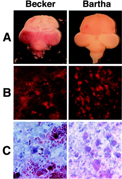
FIG. 1.
PRV infection in the chicken embryo eye model. (A) Dorsal view of whole brains 72 h after IO injection with 105 PFU in 1 μl of either the virulent Becker (left) or the attenuated Bartha (right) strain of PRV. (B) Serial cryostat sections (14-μm thick) of Becker-infected (left) or Bartha-infected (right) brains 72 h after inoculation. Sections were stained with an antibody specific to gB, followed by an Alexa 568 conjugated secondary antibody. The red fluorescence indicates virus infection. (C) Serial sections corresponding to sections shown in panel B were stained with H&E.

