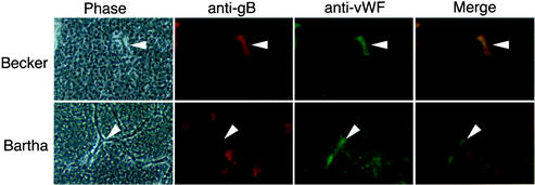
FIG. 4.
Becker, but not Bartha, is able to infect endothelial cells. Becker- and Bartha-infected brains were harvested and sectioned 72 h after infection. Sections were stained with antibodies specific to gB and vWF, an endothelial cell marker, followed by Alexa 568 and Alexa 488 conjugated secondary antibodies. The panels show phase-contrast images of the brain sections (Phase), red fluorescence indicating virus-infected cells (anti-gB), green fluorescence indicating endothelial cells (anti-vWF), and merged images of the red- and green-fluorescent panels (Merge). Arrowheads indicate blood vessels within the brain tissue.

