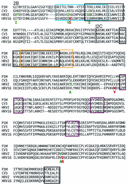
FIG. 7.
Alignment of the 2BC proteins of poliovirus, coxsackievirus, HRV2, and HRV16. Most structure-function information on 2BC comes from studies on poliovirus and coxsackievirus, and these domains were extrapolated to the rhinovirus proteins. Proteins 2B and 2C are labeled, and the cleavage site between the two is marked with a line. Outlined in purple are nucleoside triphosphate-binding motifs (36, 45). RNA-binding regions are outlined in blue (35), and membrane-binding regions are outlined in black (14, 17, 18, 44). Regions binding both membranes and RNA are outlined in orange. In the HRV16 sequence, red letters indicate amino acids that have changed in the mouse-adapted strain. In the HRV2 sequence, green letters indicate amino acids that have changed in the mouse-adapted strain. The amino acid in the adapted strain is shown below the parental sequence.

