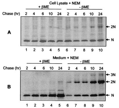
FIG. 2.
NEM-resistant disulfide bond formation between PRRSV N proteins. PRRSV-infected cells were pulse-labeled for 30 min with 150 μCi of [35S]methionine/ml and chased for the indicated times, after which the cells were solubilized in the presence of 20 mM NEM. After 2, 4, 6, 10, and 24 h of chase, the medium was collected and the virus was pelleted by centrifugation at 35,000 rpm (model XL-90; Beckman) for 2 h through a 20% sucrose cushion. The virus pellet was solubilized in the presence of 20 mM NEM. The N protein was immunoprecipitated from cell lysates (A) and purified virus particles (B), separated by SDS-PAGE under reducing (+βME) (lanes 1 to 5) or nonreducing (−βME) (lanes 6 to 10) conditions, and visualized using a PhosphorImager. The arrows indicate monomers (N), dimers (2N), and trimers (3N) of the N protein.

