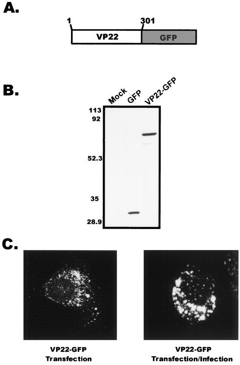
FIG. 1.
Expression of VP22 within A7 cells. (A) Construction of VP22-GFP. VP22 from the HSV-1 KOS genome was cloned as a C-terminal fusion to the GFP protein. (B) Western blot analysis. A7 melanoma cells were transfected with the indicated plasmids, and cell lysates were separated by SDS-PAGE. Proteins transferred to nitrocellulose membranes were probed with a polyclonal antibody specific for GFP. (C) Subcellular localization of VP22. Plasmid DNA encoding VP22-GFP was transfected into A7 cells as in the experiment for which results are shown in panel B, and localization of the VP22-GFP chimera was observed in the absence or presence of HSV-1 infection. Fluorescence was visualized by live-cell confocal microscopy at approximately 32 h posttransfection (transfection only) or 32 h posttransfection-infection (24 h posttransfection plus 8 h postinfection).

