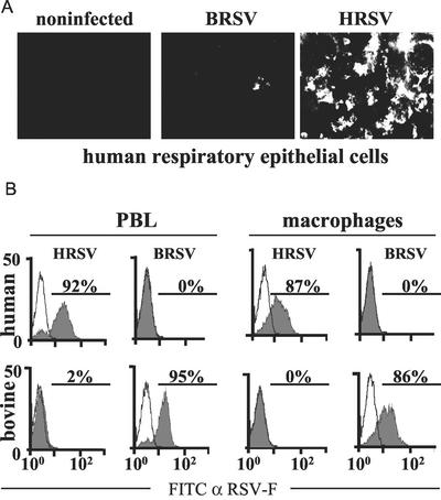
FIG. 2.
Species-specific infection of primary cells by HRSV and BRSV. (A) Monolayers of differentiated human respiratory epithelial cells were infected with the indicated viruses. Infection was visualized after 4 days by indirect immunostaining with an RSV F-specific antibody. (B) PBLs or macrophages of the indicated origin were infected with BRSV or HRSV as indicated (filled curves) or mock infected (open curves). The percentage of infected cells was determined by surface staining of RSV F with a monoclonal antibody and FACS after 6 days of infection. Results from one representative experiment of three are shown. FITC, fluorescein isothiocyanate; α-RSV-F, anti-RSV-F.

