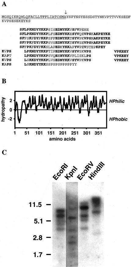
Figure 1.
Peptide structure of LeExt1 and genomic DNA gel-blot analysis. A, Deduced LeExt1 amino acid sequence. Repetitive amino acid units (indicated in bold) are arranged to emphasize various amino acid repeat units and their periodicity. The signal peptide is underlined and the putative cleavage site is marked with an arrow. B, Hydropathy plot of LeExt1 polypeptide. Hydrophilicity and hydrophobicity values are indicated at left. C, Genomic DNA was digested with the designated restriction enzymes. The LeExt1 cDNA was used as a radioactive probe. The positions of DNA marker fragments and their lengths in kb are indicated at left.

